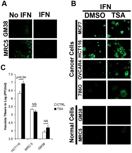
Figure 3. TSA enhances vaccinia in presence of IFN in cancer cells.
(A) Normal cells GM38 and MRC-5 were treated or not with 200 IU/ml of IFN for 16 hrs. Cells were then infected with VVdd at MOI 0.001 and fluorescent pictures were taken after 72 h. (B) Cells were pre-treated for 3 h with TSA (0.04 µM) and then 200 IU/ml of IFN for 16 h. Cells were subsequently infected with VVdd at MOI 0.001 and fluorescent pictures were taken after 72 h. (C) Cells were treated as in (B) but samples were collected after 72 h for tittering on U2OS cells. Error bars indicate the standard error. NS stands for non-significant (n = 3, ANOVA).

