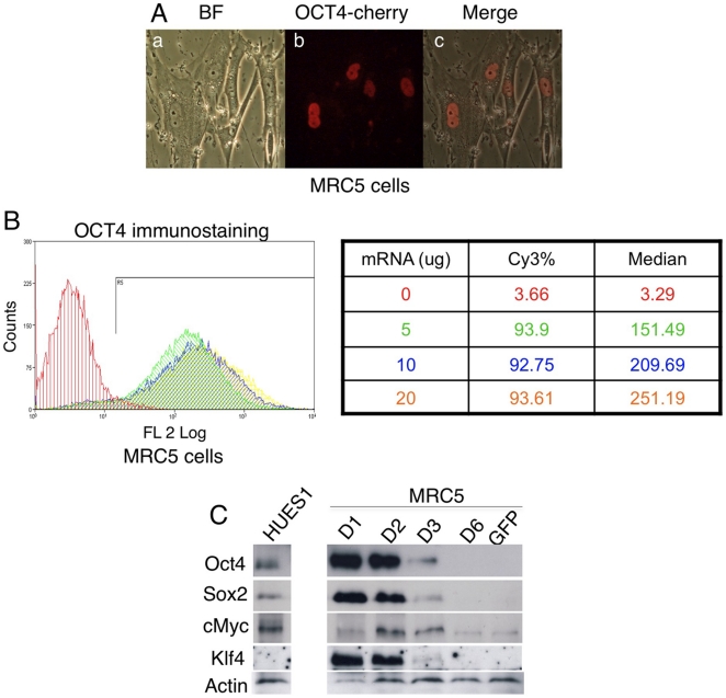
Figure 3. Protein expression following mRNA transfection.
(A) OCT4-RFP localizes into nucleus in fibroblast cells. (B) FACS analysis of OCT4 protein expression 24 hrs after mRNA microporation. Cy3 conjugated secondary antibody was used. (C)Western blot showing corresponding protein expression in 106 MRC5 cells transfected with OCT4 (17 µg), SOX2 (10 µg), cMYC (6 µg), KLF4 (6.5 µg), SV40LT (3.5 µg). The negative control is GFP mRNA transfected MRC5 cells. HUES1 is human ES cells.

