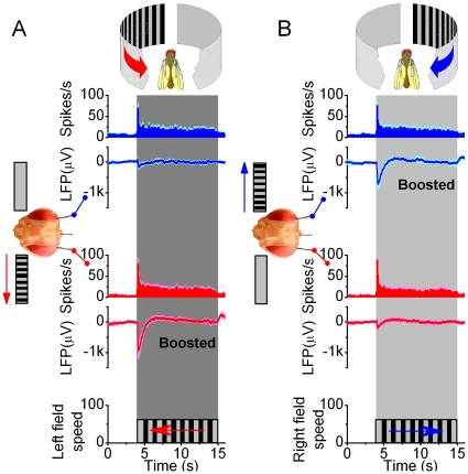
Figure 4. Brain activity increases on the side facing the motion stimulus.
Local field potentials (LFPs) in the left and right optic lobes of resting Drosophila are enhanced on the side of the moving scene (black and white stripes), whereas the firing patterns show that unilateral visual motion is processed bilaterally in the brain. (A) Neurons in both the right (blue traces) and left (red traces) optic lobes respond simultaneously and adapt rapidly to left motion; this transiently increases their firing rates, amplifying the LFPs. Peak rates: 69.6±29.0 and 79.0±38.0 spikes/s (mean ± SD; right and left electrode, respectively) show no statistical difference, whilst left LFPs are always larger (p = 0.006; ANOVA, one-way Bonferreoni test). (B) Similarly, neurons in both optic lobes respond to right motion. Peak rates: 75.3±28.7 and 87.9±40.4 spikes/s (mean ± SD; right and left electrode, respectively) do not differ statistically, but the right LFPs are always larger (p = 0.012, ANOVA, one-way Bonferreoni test). Without motion stimulus the activity is low: 5.2±1.3 spikes/s (mean ± SD; n = 12). The strong motion-sensitivity suggests that the electrodes reside in the lobula plates. Scenes were separately moved for 6–20 times on either side with 5–10 s interstimulus periods; means ± SEMs shown, n = 6 flies.

