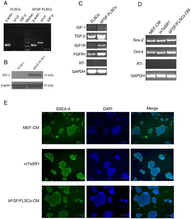
Figure 7. H9 hES cells could be maintained feeder-free with bFGF-hFLSCs-CM.
(A): RT-PCR analysis of bFGF and IGF-2 gene expression in hFLSCs and bFGF-hFLSCs. (B): Western blotting analysis of IGF-2 gene expression in hFLSCs and bFGF-hFLSCs. (C): RT-PCR analysis of bFGF and IGF-2 related gene expression in hFLSCs and bFGF-hFLSCs. (D): RT-PCR analysis of H9 hES cells grown on MEF-CM, mTeSR1 and bFGF-hFLSCs-CM for 10 passages (about 70 days). (E): Immunophenotypic characterization of H9 hES cells maintained feeder-free with MEF-CM, mTeSR1 and bFGF-hFLSCs-CM for 10 passages (about 70 days). Nuclei were stained with DAPI (blue). Bars: 250 µm.

