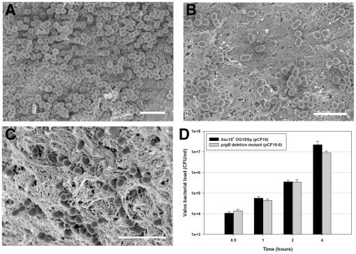
Figure 3. Asc10+ OG1SSp (pCF10) colonizes valve tissue more heavily than an Asc10− prgB deletion mutant.
(A, B, C) Valve tissue infected with Asc10+ OG1SSp (pCF10) (panel A, B) and Asc10− prgB deletion mutant strains (OG1SSp [pCF10-8]; C), as analyzed by scanning electron microscopy. Note the density of Asc10+ OG1SSp (pCF10) cells colonizing the valve (A), though not all areas of the valve were colonized as heavily, as shown in part B. In contrast, the prgB deletion mutant bound in single cells or short chains; all images were taken at 4 h post-infection. Scale bar: A, B, C = 3 µm. (D) Porcine aortic, tricuspid, and mitral valves were infected with E. faecalis strains carrying wild-type pCF10 and the prgB deletion derivative pCF10-8 for 0.5, 1.0, 2.0 and 4.0 h. Valves were washed and homogenized, and adherent bacteria were quantified by plating onto agar. The data shown are a compilation of at least three experiments, each with valve sections from a different heart.

