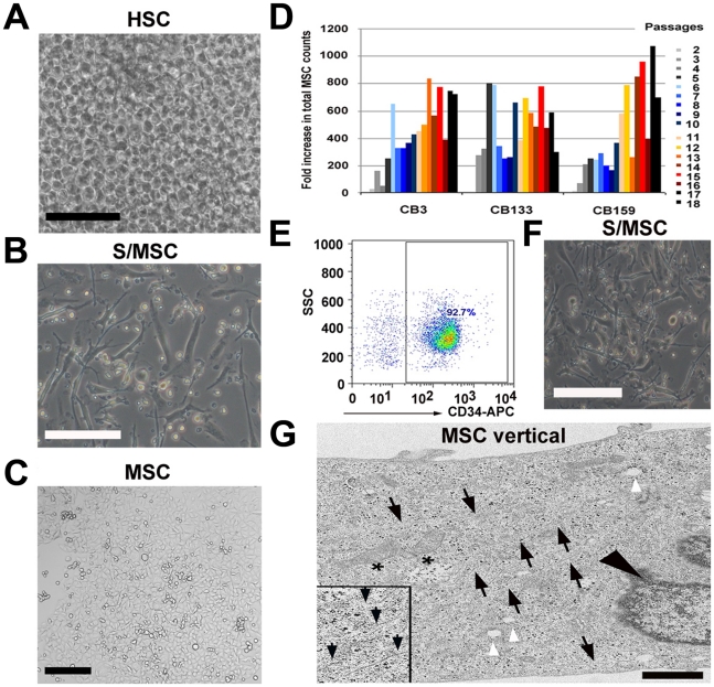
Figure 1. Umbilical cord blood stromal/adherent MSC generated during the expansion of haematopoietic stem cells in stroma-free liquid culture.
(A) Confluent growth of HSC from cultured UCB-derived MNC after 14 days expansion under stroma-free culture condition D7. Viable cell count was determined weekly (scale bar: 150 µm). (B) Adherent stromal cells after removal of non-adherent haematopietic cells and further cultivation in MesenCult with added supplements and 5 ng/ml FGF-β for an additional 14 days (scale bar: 150 µm). (C) Morphology of expanded UCB-derived MSC after 165 days/P11 in culture. After passage 1, cells were transferred from 24-well plate to 25 cm2 culture flasks and plated at a density of 80–400 cells/cm2 in MesenCult with added supplements and 5 ng/ml FGF-β. At 80% confluency, the cells were passaged again (scale bar: 100 µm). (D) Total MSC counts of 3 independent human cord blood units (CB3, CB133, CB159) performed from P2 through P18. Different colors depict different passages (2–18). Fold increase was measured by dividing the total MSC count by the starting cell number, which was 10′000 cells/25 cm2 tissue flask. (E) Percentage CD34+ selected cells using autoMACS CD34+ magnetic beads as determined by flow cytometry. (F) Adherent stromal cells generated from CD34+ selected cells expanded in D7 culture condition for 14 days. Non-adherent haematopoietic cells were removed and adherent cells were cultivated in MesenCult with added supplements and 5 ng/ml FGF-β for an additional 14 days (scale bar: 150 µm). (G) Electron microscopy (EM; vertical section) of a stromal/MSC at 198 days/P14 in culture. White arrowheads point to vacuoles and black arrowhead depicts the nucleus. Asterisks show irregular mitochondria and black arrows point to various filaments (inset, black arrows) characteristic for stromal cells. Black dots within the cytoplasm represent ribosomes (scale bar: 1 µm).

