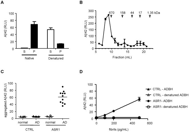
Figure 2. MPA detection of a conformational epitope in Aβ aggregates from AD brain homogenates (ADBH).
A. ADBH was centrifuged at 134,000g for 1 hr, and denatured Aβ42 in the supernatant (S) and pellet (P) fractions was detected by ELISA. B. ADBH was fractionated by size exclusion chromatography and Aβ42 was detected by ELISA. C. 75 nl of normal (open symbols) or AD (closed symbols) brain homogenate was subjected to the MPA using control (triangles) or ASR1-coated (circles) beads. D. Aβ aggregates from an ADBH were examined by the MPA with (open symbols) or without (filled symbols) a pretreatment with 5.4 M guanidine thiocyanate using control (triangles) or ASR1-coated (circles) beads. Error bars represent the standard deviation of triplicate reactions.

