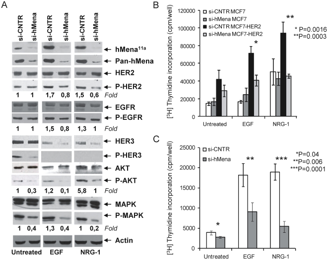
Figure 5. hMena knock-down affects HER2 signalling and inhibits the EGF/NRG1-mediated mitogenic effects in MCF7-HER2 cells.
A. Western blot analysis of MCF7-HER2 breast cancer cell line after 72 h transfection with control and hMena/hMena11a -specific siRNAs, untreated or treated with EGF (100 ng/ml) or NRG1 (10 ng/ml) for the last 24 h of transfection. hMena/hMena11a expression (evaluated by pan-hMena and hMena11a specific antibodies) and phosphorylation status of HER2, EGFR, HER3, AKT and MAPK (p44/42) from whole cell lysates were assessed. Membranes were sequentially stripped and reprobed with the indicated total and phospho-specific antibodies. Densitometric quantitation of anti-P-HER2, anti-P-AKT and anti-P-MAPK immunoreactivity was determined by Quantity One software (Biorad) and normalized in comparison with the Actin immunoreactivity. Densitometric quantitation of anti-P-HER3 immunoreactivity was not determined due to the fact that the total HER3 expression level is not unchanged following treatments. B–C. Silencing of hMena/hMena11a reduces EGF and NRG1-mediated cell proliferation of HER2 overexpressing MCF7-HER2 (B) and MDA-MB-361 (C) cell lines, but has no significant effect in MCF7 cells (B). Proliferation assays were conducted 72 h after the siRNA transfection by measuring [3H]thymidine incorporation as described in Materials and Methods. Si-CNTR: MCF7, MCF7-HER2 and MDA-MB-361 cells transfected with non-targeting siRNA; Si-hMena: MCF7, MCF7-HER2 and MDA-MB-361 cells transfected with specific hMena/hMena11a siRNA. Histograms represent the mean of three different experiments. Bars, Standard deviations. P, according to Student's t test (two tailed).

