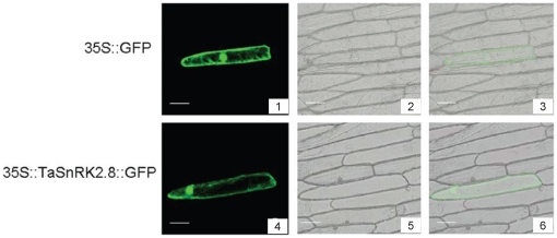
Figure 3. Subcellular localization of TaSnRK2.8 in onion epidermal cells.
Cells were bombarded with constructs carrying GFP or TaSnRK2.8-GFP as described in materials and methods. GFP and TaSnRK2.8-GFP fusion proteins were transiently expressed under control of the CaMV 35S promoter in onion epidermal cells and observed with a laser scanning confocal microscope. Images were taken in dark field for green fluorescence (1, 4). The cell outline (2, 5) and the combination (3, 6) were photographed in bright field. Scale bar = 100 µm. Each construct was bombarded into at least 30 onion epidermal cells.

