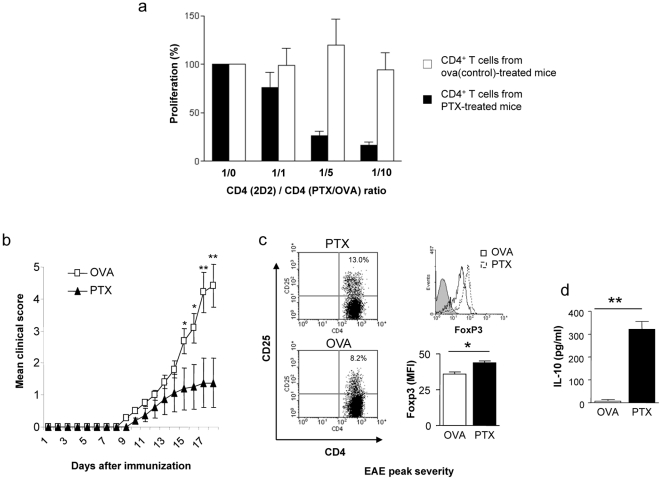
Figure 5. C57Bl/6 mice received weekly i.v. injections with 300 ng PTx or OVA 323–339 (control) in PBS.
After 4 weeks, splenic CD4+ T cells were purified (3 mice/group) and co-cultured with 2×10⁴ dendritic cells and 4×10⁴ MOG p35-55 specific T cells from TCR transgenic (2D2) mice in the presence of 20 µg/ml MOG p35-55. Proliferation at the indicated ratio of MOG p35-55 specific CD4+ T cells (2D2)/CD4+ T cells from PTx-/ova-treated mice (PTx/ova) (1/1, 1/5, 1/10) is shown as percentage of the proliferative response without CD4+ T cells from PTx-/ova-treated mice (ratio 1/0) (a). After 10 weeks, EAE was induced with MOG p35-55 (8 mice/group). PTx pre-treatment significantly ameliorated disease severity (b). At the peak of clinical EAE severity, the frequency of CD4+CD25+FoxP3+ Treg and serum level of IL-10 was evaluated. Clinical benefit of PTx-pretreated mice was associated with expansion of FoxP3+ Treg cells (c; left panels: indicated is the percentage of CD4+CD25+ within all CD4+ T cells; right upper panel: FoxP3 expression of CD4+CD25+ cells; right lower panel: mean FoxP3-FITC fluorescence intensity of CD4+CD25+ cells) and elevated serum levels for IL-10 (d) (*indicates p<0.05, ** indicates p<0.001; representative for two separate experiments).

