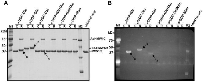
Figure 2. Glycosylation of HMW1ct by ApHMW1C.
To define the donor substrate specificity of ApHMW1C, glycosylation reactions were carried out in the reaction buffer with (R-lanes) or without (C-lanes) ApHMW1C using different UDP (or GDP) activated sugars. HMW1ct (without fusion tag) was used as the acceptor protein (lanes 1, and 3 to 6). As a control, His-tagged HMW1ct (His-HMW1ct) was also tested in a reaction with UDP-glucose as the donor sugar (lanes 2). (A) After the glycosylation reactions, samples were separated by SDS-PAGE, and the gel was stained with Coomassie Blue. (B) In parallel, a duplicate gel was transferred to a PVDF membrane and subjected to a detection reaction using the GlycoProfile III Fluorescent Glycoprotein Detection kit (Sigma). Glycosylated HMW1ct proteins are indicated by arrows: ‘a’ and ‘c’ are glycosylated HMW1ct reacted with UDP-glucose and UDP-galactose, respectively, and ‘b’ is glycosylated His-HMW1ct reacted with UDP-glucose. The lanes labeled “M1,” “M2,” and “HMW1ct only” indicate pre-staining protein markers (Precision Plus Protein Standards, Bio-Rad), glycosylated protein markers (ProteoProfile PTM Marker, Sigma), and HMW1ct only as a control, respectively.

