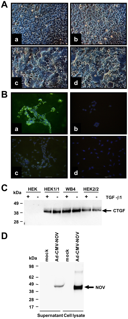
Figure 1. Establishing of devices for over-expressing CCN proteins.
(A) Light microscopic images of parental HEK cells (a), Flp-In-293 (b) and stable transfected WB4 (c) and HEK1/1 (d) clones. (B) Immunostaining for CCN2/CTGF of stable transfected WB4 (a) and parental cells (c). Staining with an isotype control (b, d) served as control. Nuclei were counterstained with propidium iode. (C) CCN2/CTGF expression in stable transfected clones and parental cells was tested in Western Blot in cells stimulated with TGF-β1 or untreated cells. (D) Expression of CCN3/NOV was analysed in extracts and supernatants of mock- or Ad-CMV-NOV-infected COS-7 cells.

