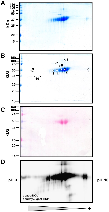
Figure 6. 2D-SDS-PAGE, trypsin in-gel digest, ESI-TOF/MS and sequence identification of protein spots from recombinant CCN3/NOV.
(A) 2D-SDS-PAGE of purified recombinant CCN3/NOV. The protein was first separated in its native state by isoelectric focussing in a pH gradient ranging from 3 to 10 and later by their protein mass in a denaturing SDS-PAGE. The final gel was stained with Coomassie Brilliant Blue. (B) Indicated spots (spot 1 to 10) were subjected to trypsin in gel digest. (C) A parallel gel of (A) was prepared and transferred without prior Coomassie stain to a Protran membrane. The membrane was stained with Ponceau S. (D) The membrane (C) was then probed with a CCN3/NOV-specific antibody.

