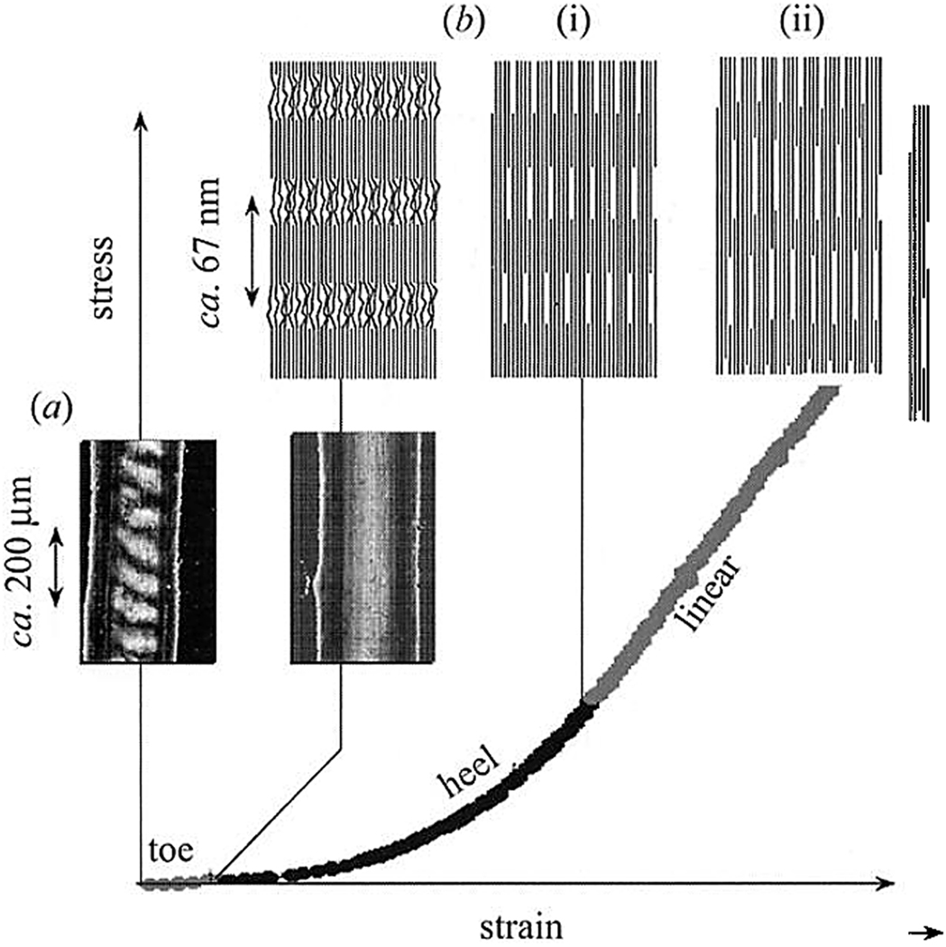
Figure 11.
Stress-strain curve of parallel arrays of collagen fibrils as found in tendons can be divided into several regions, corresponding to different strain mechanisms. (a) In the toe region, the strain is due to the straightening of the macroscopic crimp in the tissue. (b(i)) In the hill region the strain is due to the lateral alignment of the collagen molecules inside the fibrils. (b(ii)_) In the linear region the strain is due to the sliding of the collagen molecules along each other, in this region the D-spacing of collagen fibrils increases by up to 10%. The figure is reprinted with permission from reference 191.

