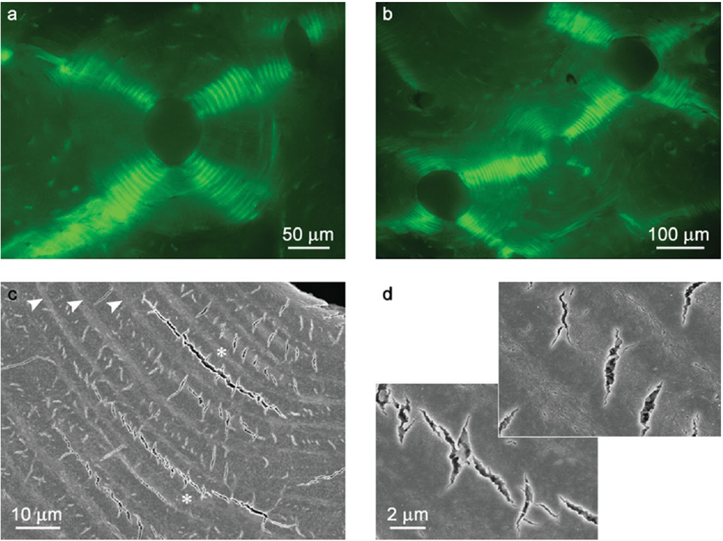
Figure 16.
Crack propagation in the osteonal bone under compression a) Epi-fluorescence image showing four groups of arc-shaped circumferential microcracks (bright green) arranged in the quasiorthogonal pattern; b) Epi-fluorescence image showing the crack propagation across neighboring osteons. The cracking of the central osteon is transfered to the osteons to the lower left and upper right; c) SEM micrograph showing arc-shaped microcracks. d) Closer observations of (c) (asterisks) showing the short micro-radial cracks in the thick lamellae and a circumferential microcrack. The figure is reprinted with permission from reference 202.

