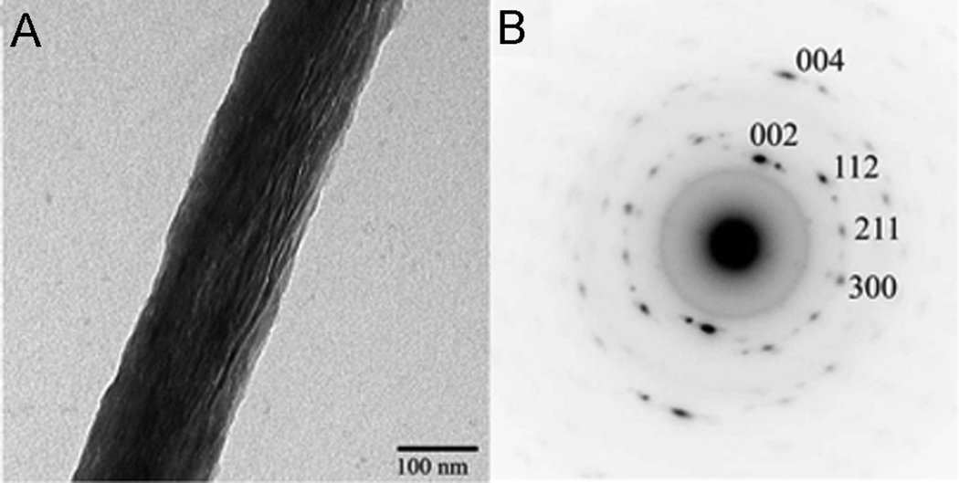
Figure 9.
A. TEM micrograph of collagen fibril mineralized in vitro in the presence of polyaspartic acid. Note that the mineral particles are aligned along the fibril axis. B. Electron diffraction pattern of the mineralized fibril in Figure 9A.; c-axes of the crystals are co-aligned with the fibril axis. The figure is reprinted with permission from reference 105.

