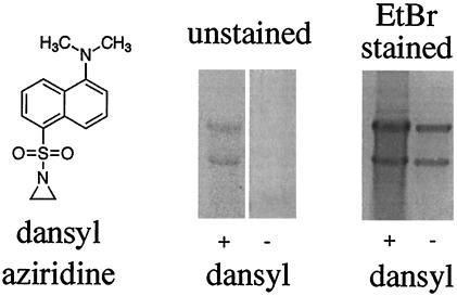
Figure 3.
Agarose gel electrophoresis of N-dansylaziridine-modified and -unmodified viral RNA. RNA was extracted from N-dansylaziridine-treated FHV and an untreated control sample by using phenol-chloroform. The extracted material was ethanol-precipitated and electrophoresed through a nondenaturing 1% agarose gel. (Left) Structure of N-dansylaziridine. (Center) After electrophoresis, the gel was placed on a transilluminator and viewed by using a 300-nm light source. Nucleic acids representing FHV RNA1 (3.1 kb, upper band) and RNA2 (1.4 kb, lower band) were visible for the sample treated with N-dansylaziridine, but not the control. (Right) The gel was stained with ethidium bromide for 30 min and viewed again with a transilluminator. RNA in both lanes is now visible.

