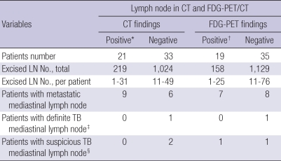
Table 2.
Pathological diagnosis of the dissected lymph nodes with positive findings on CT and FDG-PET/CT among patients who had undergone both CT and FDG-PET/CT
*The presence of lymph nodes ≥ 1 cm in their smallest diameter was considered to be positive on CT; †The presence of lymph nodes with a maximum SUV ≥ 3.0 was considered to be positive on FDG-PET/CT; ‡Granulomas and acid-fast bacilli were observed on microscopic evaluation in one patient only; §Only granulomas were observed on microscopic evaluation in two patients. CT, computed tomography; FDG-PET, fluorodeoxyglucose-positron emission tomography; LN, lymph node; SUV, standardized uptake value; TB, tuberculosis.

