Figure 7.
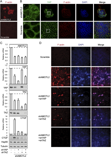
AMOTL2 knockdown induces cell transformation in MDCK cells in a YAP- and TAZ-dependent manner. (A) AMOTL2 knockdown in MDCK cells induces foci formation. Scramble and AMOTL2 knockdown MDCK cells were established by lentiviral infection of MDCK cells with shRNA constructs. Cells were then cultured onto transwell membranes at a density of 1 million cells per 24-mm insert. After 5 d, cell foci were visualized by rhodamine-phalloidin staining. (B) AMOTL2 knockdown-induced cell foci have elevated nuclear YAP levels. Cells the same as those in A were fixed and stained with anti-YAP (from Dr. Marius Sudol's laboratory) to visualize YAP localization, and with rhodamine-phalloidin to show cell boundaries. DAPI labels cell nuclei. Images of cells in shAMOTL2-induced foci above the monolayer (top panels) and of cells in the monolayer of control cells (bottom panels) are shown. Panels 1 and 2 represent insets from YAP staining as indicated. (C) Knockdown of YAP and TAZ blocks AMOTL2 knockdown-induced CTGF expression. MDCK cells infected with indicated shRNA constructs were established. Levels of indicated mRNAs and proteins were analyzed by quantitative RT–PCR and Western blots, respectively. Anti-tubulin and anti-Hsp90 Western blots were used as loading controls. (D) Knockdown of YAP and TAZ blocks AMOTL2 knockdown-induced foci formation. Cells the same as those in C were tested in foci formation assays following the procedures in A. Cell foci were visualized by both rhodamine-phalloidin staining of F-actin and DAPI staining of cell nuclei.

