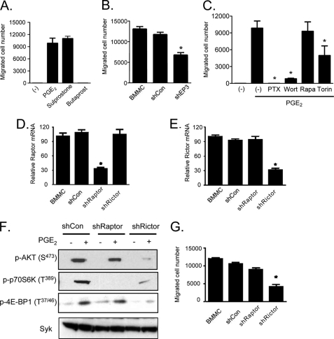
FIGURE 2.
PGE2-induced chemotaxis is mediated by mTORC2. A, cytokine-deprived BMMCs were prepared, and the chemotaxis assay was performed in response to 100 nm of PGE2, sulprostone, or butaprost. B, BMMCs were transduced with lentivirus expressing shRNA for EP3, and the chemotaxis assay was performed in response to 100 nm of PGE2. C, BMMCs were preincubated with the indicated inhibitors (wortmannin (Wort), rapamycin (Rapa), or Torin (100 nm, 30 min) or PTX (1 μg/ml, 4 h)), then washed, and used for the PGE2 (100 nm)-induced chemotaxis assay as described under “Experimental Procedures.” D–G, BMMCs were transduced with lentivirus expressing control shRNA (shCon), shRNA for raptor (shRaptor), or shRNA for rictor (shRictor) as described under “Experimental Procedures.” To establish knockdown of raptor or rictor, cDNA was prepared from the cells, and real time PCR was performed using commercial TaqMan raptor or rictor probes (D and E). To confirm the knockdown, raptor or rictor's downstream molecule phosphorylation was tested. The cells were stimulated with PGE2 (100 nm) for 10 min, and protein lysates were subjected to Western blotting (F). After confirming raptor and rictor knockdown, the PGE2-induced chemotaxis assay (G) was performed. The results are the means ± S.E. of three separate experiments. *, p < 0.05 for comparison with PGE2 alone stimulation (C) or with shCon (B, D, E, and G) by Student's t test.

