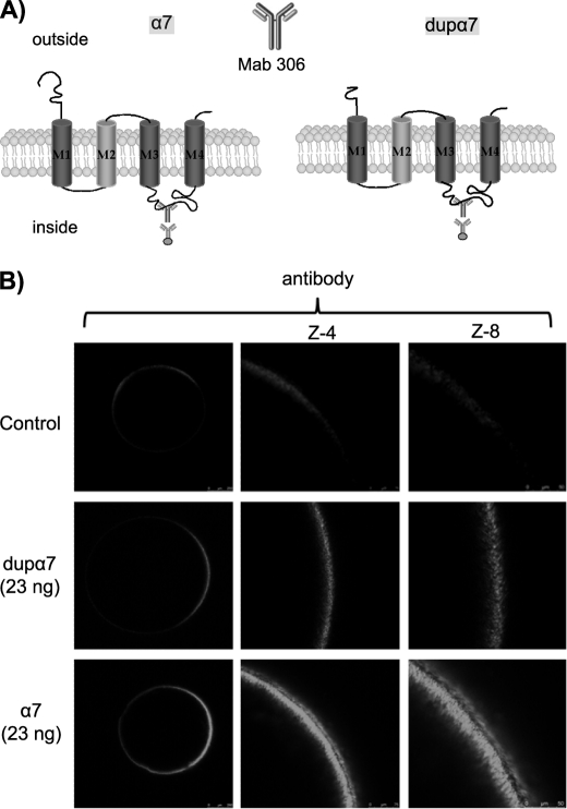
FIGURE 2.
Confocal images of oocytes expressing dupα7 and α7 subunits. A, diagram of tertiary structures of α7 and dupα7 proteins; the Mab306 antibody binding region is also indicated. B, left column shows overall confocal (Z-scan) images of three typical intact oocytes (out of five), noninjected (control), or injected with dupα7 or α7 mRNA and immunostained with the antibody. Central and right columns show, at a higher magnification, the fluorescent signal at the animal surface in the three oocytes.

