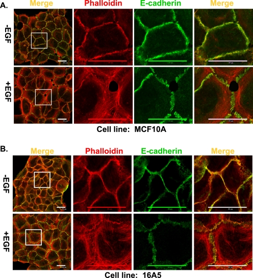
FIGURE 1.
EGF stimulation of MECs results in remodeling of junctional actin cytoskeleton and disruption of AJs. MCF10A cells (A) and 16A5 cells (B) were grown without EGF for 4 days and stimulated with EGF (3 ng/ml) for 12 h. The cells were immunostained and scanned with confocal microscopy at the subapical plane for E-cadherin (green) or actin (phalloidin; red). Both the whole cell colony image and the high resolution image of the indicated areas in the colonies are presented (scale bar, 20 μm).

