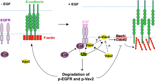
FIGURE 10.
Model of Cbl-dependent stabilization of AJs in EGF-stimulated MECs through negative regulation of EGFR and Vav2 signaling. MECs form AJs by E-cadherin clustering, which is facilitated by tight circumferential actin cables. EGFR phosphorylates Vav2 that translocates to the cell membrane at the cell-cell junctions and activates Rac1/Cdc42 that reorganizes junctional actin, resulting in de-clustering of E-cadherin and disruption of cell-cell adhesions. Attenuation of Rac1/Cdc42 activation is partially achieved by Cbl-mediated ubiquitinylation of EGFR resulting in attenuation of phosphorylated Vav2. Cbl can additionally lead to ubiquitinylation (Ub) and degradation of p-Vav2 resulting in its removal from the junctional membrane, therefore temporally and spatially restricting Rac1/Cdc42 activation and reorganization of junctional actin cytoskeleton. This attenuation is hypothesized to serve as a physiological barrier to prevent excessive reorganization of junctional actin cytoskeleton and complete loss of AJs during EGF-induced MEC remodeling process.

