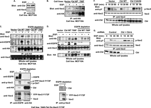
FIGURE 5.
EGF-induced Cbl-Vav2 association and Cbl-dependent attenuation of Vav2 phosphorylation. A, growth factor-starved MCF10A cells were stimulated with EGF (100 ng/ml) for 10 min, and anti-Cbl IPs from whole cell lysates were immunoblotted for Vav2 and Cbl. B, MCF10A cells with stable overexpression of HA-tagged wild type Cbl or Cbl-Y700F were stimulated with EGF (100 ng/ml) for 10 min and anti-HA IPs immunoblotted for Vav2. C, equal amount of cell lysates from the cells with vector or wild type Cbl overexpression and one-third amount of cell lysates from cells with Cbl-Y700F expression prepared under the same experimental conditions as described in B were subjected to IP (1st to 6th lanes) with anti-HA Abs as such or after immunodepletion of EGFR before (7th to 10th lanes) and then immunoblotted for Vav2, EGFR, and HA. D, equal amounts of cell lysates from vector-, wild type Cbl-, or Cbl-Y700F-overexpressing cells under the same experimental conditions as described in B were subjected to IP with anti-Vav2 Abs (1st to 6th lanes) or immunodepleted of EGFR before IP with anti-HA Abs (7th to 10th lanes) followed by immunoblotting for HA, EGFR, and Vav2. E, growth factor-starved and DOX-induced 16A5-Tet-On-Vav2-Y172F/HA-Cbl-expressing cells were treated with EGF (100 ng/ml) for 5 min. Equal amounts of cell lysates were subjected to serial (three times) anti-EGFR IP to immunodeplete EGFR (left panel, 1st and 2nd lanes) before IP with anti-HA (left panel, 3rd and 4th lanes) or with anti-phospho-Vav2 antibodies (right panel) and immunoblotted for EGFR, phospho-Vav2, Vav2, and HA. F, growth factor-starved MCF10A cells with stable overexpression of vector, wild type Cbl or Cbl-C3AHN mutant were stimulated with EGF (100 ng/ml) for the indicated time. Equal amounts of cell lysates were subjected to IP with anti-Vav2 Abs and immunoblotted for Tyr(P) (pY) and Vav2. Whole cell lysates were also blotted for Cbl (lowest panel). G, MCF10A cells with stable expression of control shRNAs or Cbl/Cbl-b shRNAs were stimulated with EGF (100 ng/ml). Cell lysates were subjected to IP with anti-Vav2 Abs and blotted for Tyr(P) (pY) and Vav2. Whole cell lysates were also immunoblotted for Cbl (lowest panel).

