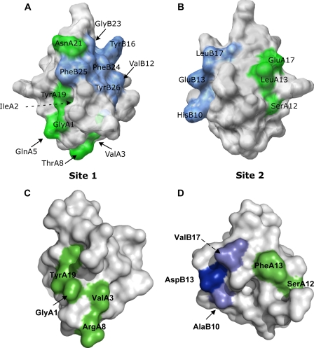
FIGURE 8.
Conservation of insulin and DILP5 binding surfaces involved in binding to the human insulin receptor. Panel A and B depict mammalian insulin binding sites 1 and 2 (70). Panels C and D show the conserved residues that are involved in mammalian insulin binding in a space-fill illustration of DILP5-DB. The residues involved in binding to the receptor are illustrated in green (A-chain residues) and blue (B-chain residues). The lighter blue color represents conservative substitutions. PDB code 9INS (mammalian insulin) and 2WFU (DILP5-DB).

