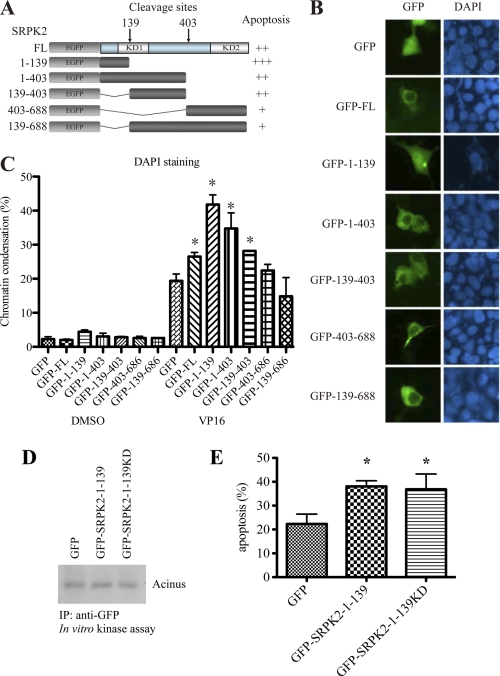
FIGURE 6.
N terminus of SRPK2 localizes to the nucleus and promotes apoptotic cell death. A, schematic representation of various SRPK2 fragments that mimic the cleaved products by caspase-3. B, subcellular localization of SRPK2 fragments. HEK293 cells were transfected with GFP-tagged SRPK2 fragments for 24 h. The cell nuclei were stained with DAPI and visualized under a fluorescent microscope. C, apoptosis quantification. HEK293 cells were transfected with GFP-tagged SRPK2 fragments for 24 h, and then treated with DMSO or VP 16 for another 16 h. The cells were stained with DAPI- and GFP-positive cells were counted for chromosome condensation. *, p < 0.05. A minimum of 300 cells were counted from 3 different fields. D, SRPK2-(1–139) and SRPK2-(1–139)KD possess no kinase activity. GFP, GFP-SRPK2-(1–139) or GFP-SRPK2-(1–139)KD was transfected into HEK293 cells and then pulled-down with anti-GFP antibody. The precipitated proteins were incubated with purified Acinus as a substrate and analyzed by in vitro kinase assay. E, quantification of apoptosis. HEK293 cells were transfected with GFP, GFP-SRPK2-(1–139) or GFP-SRPK2-(1–139)KD for 24 h and then treated with VP16 for another 16 h. The adherent and non-adherent cells were collected, mounted on the slides, and stained with DAPI. The percentages of cells with chromosome condensation were counted. *, p < 0.05. A minimum of 300 cells were counted from 3–5 different fields.

