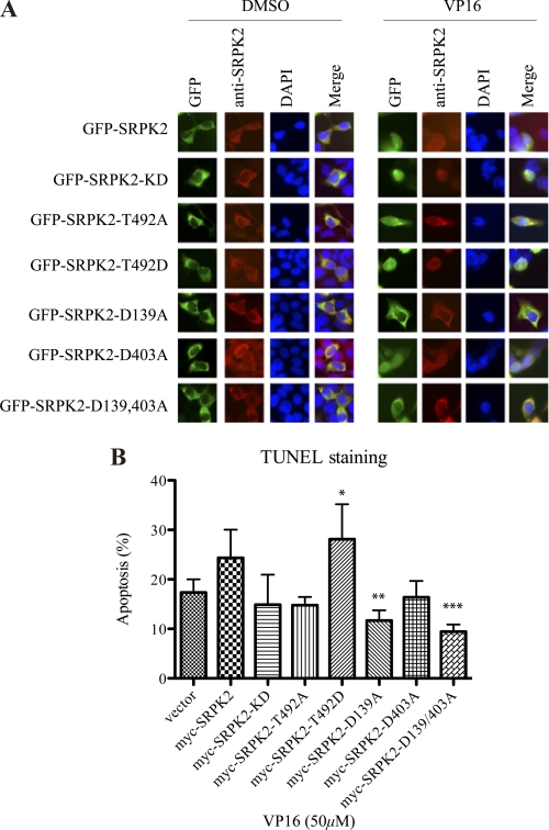
FIGURE 7.
D139A mutation inhibits the pro-apoptotic effect of SRPK2. A, subcellular localization of GFP-SRPK2 and its mutants upon VP16 treatment. HEK293 cells were transfected with GFP, GFP-tagged SRPK2WT, KD, T492A, T492D, D139A, D403A, or D139/403A for 24 h, and then treated with DMSO or VP16 for another 24 h. The cells were stained with DAPI and visualized under the fluorescent microscope. B, quantification of apoptosis. HEK293 cells were transfected with empty vector, Myc-tagged SRPK2WT, KD, T492A, T492D, D139A, D403A, or D139/403A for 24 h, and then treated with DMSO or VP16 for another 16 h. Then, the adherent and non-adherent cells were collected, mounted on the slides and stained with the TUNEL reagents and DAPI. The percentages of TUNEL-positive cells were counted. *, vector versus T492D; **, SRPK2 versus D139A; ***, SRPK2 versus D139/403A, p < 0.05. A minimum of 300 cells were counted from three different fields.

