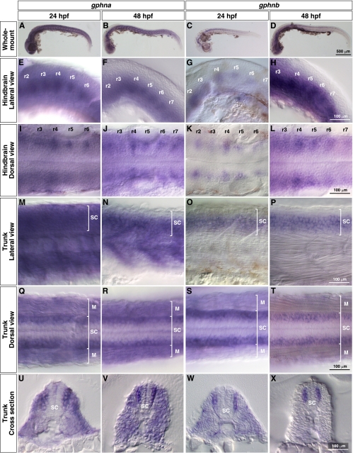
FIGURE 4.
Spatial expression of zebrafish gphna and gphnb. In situ hybridization with gphna and gphnb antisense probes. A–D, whole-mount images. Lateral views (E–H) and dorsal views (I–L) of hindbrains show gphna and gphnb expression in bilaterally located reticulospinal neurons of rhombomere segments (r2–r7). Lateral views (M–P) and dorsal views (Q–T) of trunks show gphna expression in muscles (M) and spinal cords (SC) and gphnb expression in spinal cord at 24 and 48 hpf. Cross-sections of trunks (U–X) reveal that both gphna and gphnb are expressed in lateral spinal neurons.

