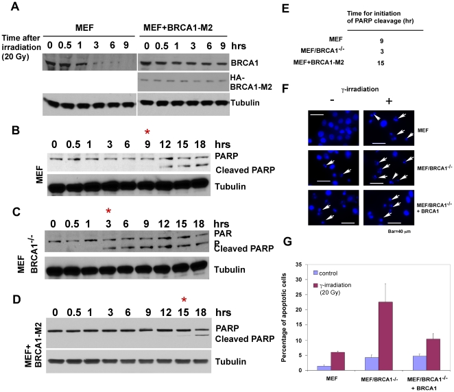
Figure 6. BRCA1 is important in modulating the onset of apoptosis in the presence of γ irradiation.
A. BRCA1 is degraded in response to γ irradiation in MEF cells. B. Data based on measurements of PARP cleavage showed that initiation of γ irradiation-induced apoptosis (at 20 Gy) was detected about nine hours after exposure to γ irradiation. C. Loss of BRCA1 significantly enhanced the onset of apoptosis as reflected by an approximate six hours upshift for PARP cleavage. D. Expression of a non-degradable BRCA1 in MEF cells delayed the onset of γ irradiation-induced apoptosis. E. Summary of time for activation of apoptosis under different background of BRCA1. F. Apoptosis was visualized by fluorescence microscopy. γ irradiation treated cells were fixed and stained with DAPI and nuclear morphology was observed. Arrow indicates apoptotic cells. G. Quantification of γ irradiation-induced apoptosis in MEF, MEF/BRCA1−/−, and MEF/BRCA1−/− + BRCA1 cells. Cells were stained with Annexin V and PI and apoptotic cells (Annexin V+/PI −) were quantified by FACS. Results are mean ± s.d. of three independent experiments (G).

