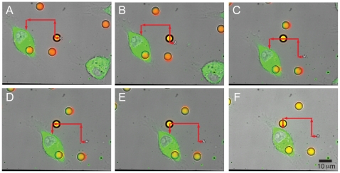
Figure 2. Trapping and positioning of polystyrene bead next to a fluorescent J774 cell.
(a–f) Combined fluorescent and brightfield images extracted from a real-time movie of a fluorescent polystyrene bead (yellow) positioned next to a fluorescent J774 cell expressing YFP-actin (green) (see Supporting Information for Video S1). The bead is trapped in a field of other fluorescent beads (orange) and another fluorescent J774 cell (also labeled green) as the stage is moved to position the cell next to the trapped bead. The red arrows indicate the deflection of the stage to direct the bead adjacent to the J774 cell, and the starting position of the trapped bead is indicated by a star (*).

