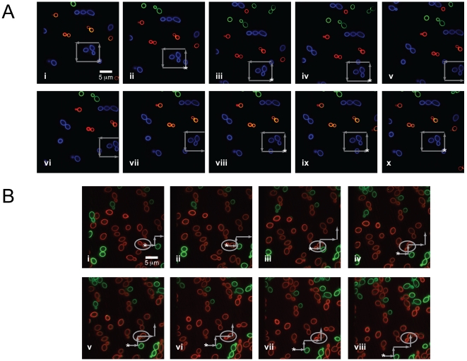
Figure 3. Fluorescence images of trapped and manipulated C. albicans.
(a) A trapped fluorescently-labeled (Alexa Fluor 488 (AF488), blue) organism in a field of other fluorescently-labeled C. albicans (AF568, green and AF647, red). The stage is moved around the trapped, blue CA particle as indicated by the gray arrows, and follows the sequence in frames i–x. The starting position of the CA is indicated by a star (*), and briefly moves out of the field of view in frames v–vii. Full length movie of trapped C. albicans can be seen in Supporting Information, Video S2. (b) Trapped C. albicans with attached, budding daughter cell (labeled with AF647, red, and highlighted in the gray circle) in a highly dense field of AF647-labeled and AF488-labeled (green) organisms. The stage is maneuvered around the red C. albicans, as indicated by the gray arrows. Frames i-viii shows the sequence of movements to position C. albicans in a different area of the stage. The starting position of the mother-daughter pair is indicated by a star (*).

