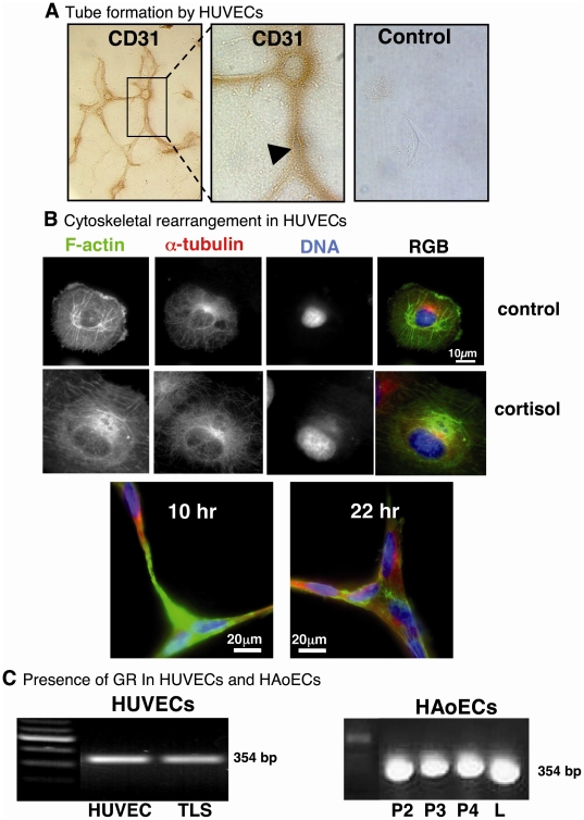
Figure 1. Formation of tube-like structures (TLS) and expression of glucocorticoid receptors (GR) in human endothelial cells.
(A) Human umbilical vein endothelial cells (HUVECs) cultured on Matrigel formed a network of tube-like structures (TLSs), after approximately 4 hrs, that retained immunoreactivity for the endothelial cell marker CD31 (original magnification ×10). At higher magnification, cell membranes were evident (arrow head), suggesting development of a lumen in the cell-cell connections (original magnification ×40). No staining was observed in negative controls lacking primary antibody. (B) HUVECs cultured on uncoated cover slips showed clearly-defined cytoskeletal components: filamentous (F)-actin (stained with phalloidin-488; green) and α-tubulin (stained with goat anti-mouse IgG Alexa Fluor 594 secondary antibody; red), and nucleus (DNA stained with 4′,6-diamidino-2-phenylindole dihydrochloride (DAPI); blue). Exposure to cortisol (600 nM; 1 hour) had no apparent effect on microtubule staining but induced a more diffuse and homogeneous distribution of F-actin throughout cell. TLSs stained after 10 hours or 22 hours in culture, consisted of adjoining, filopodia-like extensions, containing both F-actin (green) and α-tubulin (red), connecting neighbouring cells (DNA, blue). (C) GR were detected both in first passage (P1) human umbilical vein (HUVECs) and in passaged human aortic (HAoECs) endothelial cells (P2–P4), by RT-PCR (354 bp product). GR expression was maintained in HUVECs 22 hours after TLS formation. L, Liver (positive control). Negative controls included no reverse transcriptase and no cDNA (not shown).

