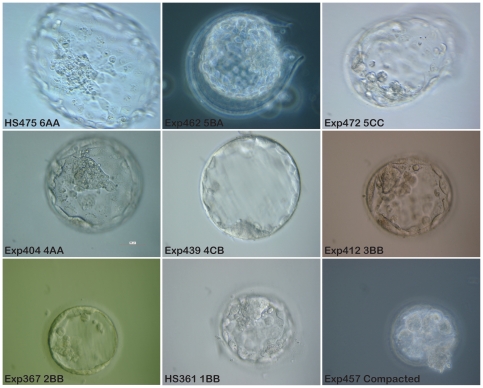
Figure 1. Photographic examples representing the different scores of blastocysts donated for stem cell derivation.
HS475. Fully hatched blastocyst with clear tightly packed ICM. This embryo generated a new hESC line, HS475. Exp462. Hatching blastocyst with average number of cells in ICM, even trophectoderm layer. Exp472. Hatching blastocyst. Trophectoderm layer is uneven and the cells in the ICM are few in number and loosely packed. Exp404. Fully expanded blastocyst with clear tightly packed ICM and even trophectoderm layer. Exp439. Fully expanded blastocyst, with thin zona pellucida. Small ICM and trophectoderm cells of different size. Exp412. Fully developed blastocoel. Zona pellucida is still thick, meaning that the blastocyst is not fully expanded. Exp367. Early blastocyst. HS361. Early blastocyst with blastocoels filling less than 50% of embryo volume. This embryo generated the hESC line HS361. Exp457. This embryo is still compacted and has not reached blastocyst stage. All pictures were taken with a 40x objective, phase contrast or DIC, scale bar 50 µm. (Exp = experiment number. HS is used as a prefix for established hESC lines at Karolinska Institutet).

