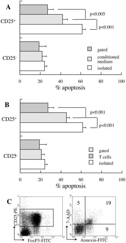
Figure 2. Soluble and cellular factors affecting susceptibility to apoptosis.
A. CD25− and CD25+ T cells isolated from new onset diabetic NOD females were incubated for 48 hours in conditioned medium from CD25− T cells stimulated with surface-bound anti CD3 and anti-CD28 antibodies (n = 3). Data are compared to corresponding measurements of isolated cells and mixed cultures. B. Spontaneous apoptosis after 48 hours of culture of isolated CD25+ T cells (n = 4) and gated subsets in mixed cultures (n = 5) following B220, GR-1 and MAC-1 depletion (n = 4). C. Equal numbers of isolated CD25− and CD25+ T cells from diabetic NOD mice were mixed for determination of apoptosis after 48 hours of culture in the CD25+ subset (gate). Data are representative of four independent incubations.

