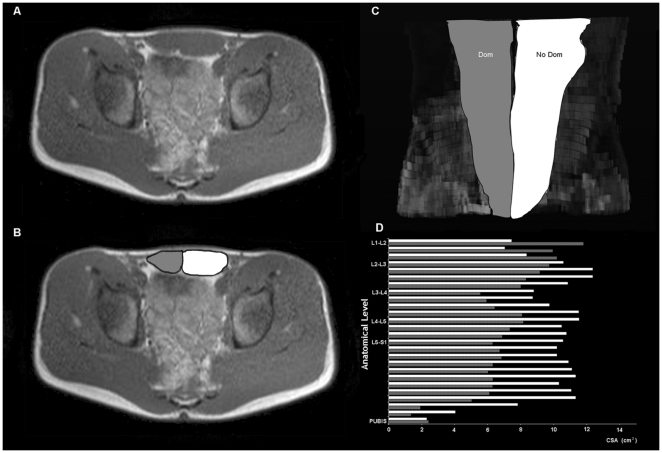
Figure 1. Digital reconstruction of rectus abdominis muscle of one right-handed professional tennis player, from magnetic resonance images (MRI).
(A) Cross sectional MRI at the umbilical level, and (B) corresponding image showing the different muscle compartments measured. (C) Digital reconstruction of rectus abdominis muscle in the coronal plane, from L1-L2 to the pubic symphysis, and (D) figure illustrating the successive cross sectional MRI measurements performed. In gray, the dominant side (Dom), in white, the non dominant side (NoDom) of rectus abdominis muscle.

