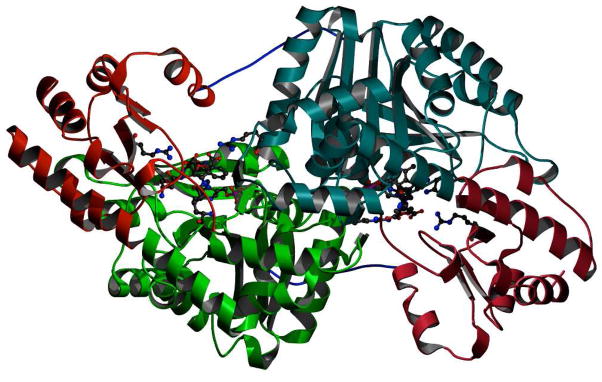
FIGURE 6.
Dimeric structure of S-ADFA inactivated L-AspAT at pH 8. The two large domains are in dark and light green respectively. The two small domains are in dark and light red. The active sites are indicated with bound inhibitors, and with catalytic and recognition residues shown arising from the domains and subunits from which they derive. The first 12 amino acids, in blue, show the interactions between the small domain of one subunit with the large domain of the other subunit; these residues are not always visible in the electron density of L-AspAT structures but are visible in these reported here.

