
Figure 2.
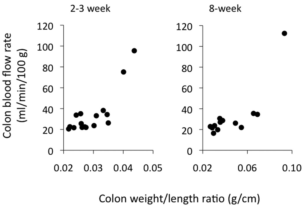
Colon blood flow rates in the IL-10−/− CD4+ ⇒ RAG−/− mice 2–3 and 8 weeks following reconstitution, as plotted vs the colon weight-to-length ratio on the x-axis. The occasional higher blood flow rates were found in the mice with higher weight-to-length ratios. Note the change in x-axis scale as the weight-to-length ratio increased with time.