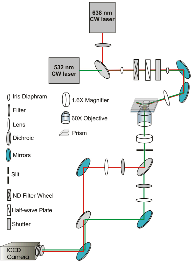
Figure 3.
Single-molecule prism-based TIRFM setup. Linearly polarized light from the 630-nm red diode laser passes through a 638 ± 10 nm clean-up filter and is reflected off a 610?-nm cutoff dichroic. 532-nm light from the Nd:YAG passes through same dichroic to join the same beam path. Light from both lasers then passes through the following components, in order: an iris diaphragm, a neutral density filter, a λ/2-wave plate for 532-nm light, a shutter, an iris diaphragm, a series of mirrors (two are shown for ease of representation; three were used during our experiments), a focusing lens, the TIR prism, the microscope slide containing the sample, the 60X objective, a 1.6X magnifier (along with additional filters, mirrors, and image transferring lenses contained within the microscope), a slit, and a 610-nm cutoff dichroic mirror. The emission from Cy3 passes through a band-pass filter, while that of Cy5 passes through a long pass filter. These separated images are projected side-by-side onto an intensified charge-coupled device (ICCD) camera.

