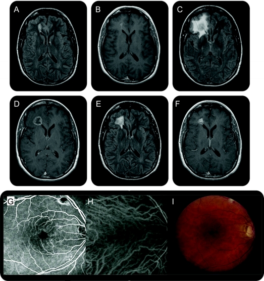
Figure Brain and retinal imaging in a patient with cerebroretinal vasculopathy
Sequential axial MRI showing an ovoid T2-hyperintense (A) and gadolinium-enhancing (B) lesion (2.1 × 1 cm), abutting the frontal horn of the right lateral ventricle, bright on both diffusion-weighted imaging (DWI) and apparent diffusion coefficient (ADC) map. At 6 months, a larger, more aggressive-appearing lesion (2 × 2 × 3 cm) with surrounding edema occupied the right frontal lobe. There was a central zone of presumed necrosis and gadolinium enhancement of the lesion rim (C, D). Diffusion imaging showed more heterogeneous signal, without the characteristic bright DWI and dark ADC signal of acute infarction. At 12 months, the lesion approximated its size on initial imaging (1.4 × 1.2 × 0.9 cm) with a persistent rim pattern of enhancement (E, F). At 18 months, the lesion further decreased in size (1.1 × 0.9 × 0.3 cm) with near resolution of the surrounding edema. Fluorescein angiography and indocyanine green with corresponding color photographs of the retina show views of the macula of the right eye (G–I). Periarteriolar narrowing and sheathing, focal leakage, telangiectasias, and cotton wool spots are present.

