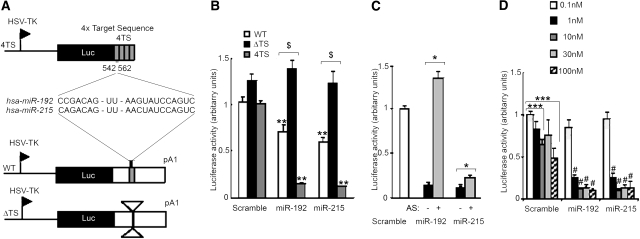
Figure 3.
miR-192 and miR-215 functionally interact with their target sequence within WNK1 3′UTR in vitro. (A) Schematic representation of the luciferase expression vectors. (B) Luciferase to β-gal chemiluminescence ratios measured when cotransfecting MDCK cells with the indicated luciferase reporter and 30 nM of either a scramble miR (Sc), miR-192, or miR-215. Cotransfection of the WT or 4TS plasmid with miR-192 or miR-215 induced a significant decrease in luciferase activity compared with the scramble (**P < 0.01). Inhibition of luciferase activity was lost in cells cotransfected with the ΔTS plasmid and any of the two miRs compared with cells cotransfected with the WT plasmid ($P < 0.0005). (C) Luciferase to ß-gal chemiluminescence ratios measured when cotransfecting MDCK cells with the 4TS reporter and 30 nM of either Sc, miR-192, or miR-215, alone or in combination with their inhibitor. The inhibition induced by the miRs was reversed by addition of their inhibitor (*P < 0.05). (D) Luciferase to ß-gal chemiluminescence ratios measured when cotransfecting MDCK cells with the 4TS reporter and increasing quantities (0.1, 1, 10, 30, or 100 nM) of either Sc, miR-192, or miR-215. Surprinsingly, luciferase activity was inhibited by cotransfection of 10 or 100 nM of Sc compared with 0.1 nM (***P < 0.005) but was significantly more suppressed by transfection of increasing concentrations of miR-192 and miR-215 (#P < 0.005 compared with the same concentration of Sc). Results are expressed as mean ± SEM, from three independent transfections realized in triplicate.

