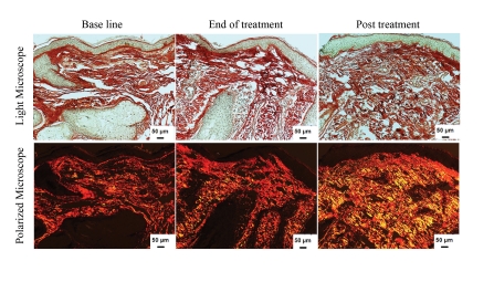
Figure 4.
Increase in newly synthesized collagen content in response to ELOS treatment. Representative examples of skin biopsies stained with picrosirius red viewed under bright field microscope (top panels) and polarized field (bottom panels). Bright field captures total collagen content. Polarized light shows a yellow-to-orange birefringence with newly synthesized collagen in yellow and total collagen in red. Note the increase in newly synthesized collagen as reflected by the yellow color after ELOS treatment.

