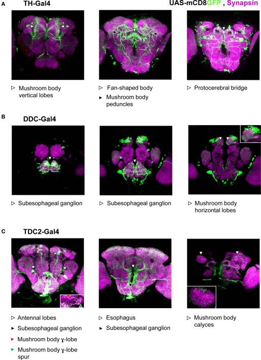
Figure 3.
Approximated patterns of Gal4 expression by the used drivers. We drove the expression of a membrane bound green fluorescent protein (mCD8GFP) using three different Gal4 drivers. Patterns of GFP-immunoreactivity (green) should approximate the respective patterns of Gal4-expression; Synapsin-immunoreactivity (magenta) shows the organization of the neuropils. We display projections of frontal optical sections of 0.9 μm, each. In each row, the leftmost panel shows the anterior-most projection; in each panel, dorsal is to the top. When driven by TH-Gal4 (A), GFP was expressed in neurons that innervate the mushroom body vertical lobes and peduncles (left and middle panels) as well as the fan-shaped body (middle panel) and the protocerebral bridge (right panel). We found no innervation of the antennal lobes or the mushroom body calyces (but see Mao and Davis, 2009). Under the control of the DDC-Gal4 driver (B), GFP was expressed in neurons that innervate the subesophageal ganglion (left and middle panels) as well as the horizontal lobes of the mushroom body (right; see also the inset). Neurons that express GFP, driven by TDC2-Gal4 (C) innervated the antennal lobes (left panel), mushroom body γ-lobes and their spurs (left panel, inset), the subesophageal ganglion (left and middle panels), the areas surrounding the esophagus (middle panel), and the mushroom body calyces (right panel; see also the inset).

