Fig. 2.

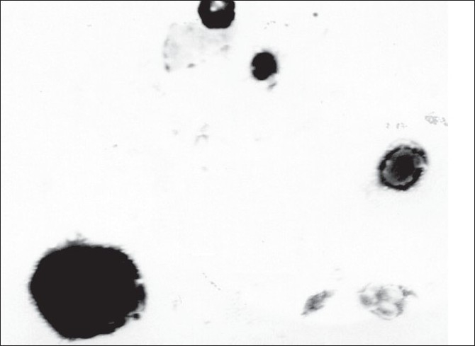
TEM photomicrograph of BDS liposomes TEM images of BDS loaded liposomes appearing as bright spheres surrounded by dark thick layer showing large internal aqueous core

TEM photomicrograph of BDS liposomes TEM images of BDS loaded liposomes appearing as bright spheres surrounded by dark thick layer showing large internal aqueous core