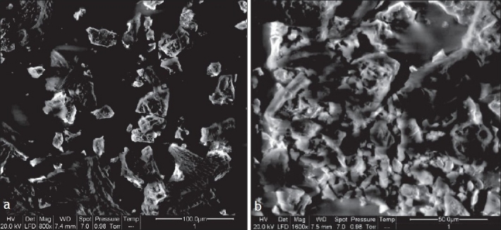
Fig. 3.

ESEM photomicrograph of freeze dried trehalose particles ESEM photomicrograph shows that freeze dried trehalose particles (a) were of irregular porous particles and freeze dried liposomes (b) appeared as aggregated particles with lipids on the surface
