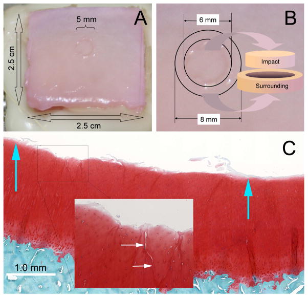
Figure 1.
Osteochondral Explant Blunt Injury Model. (A) The image shows the gross appearance of a 14J/cm2 blunt impact injury on the surface of a typical osteochondral explant. The center of the 2.5 cm × 2.5 cm explant was struck once with a 5-mm diameter platen. (B) Illustration of the method employed to harvest cartilage tissue from a traumatized explant. Six mm punches were used to harvest the impact site itself and 8 mm punches were used to harvest an adjacent annular ring of cartilage surrounding the impact site. (C) A high resolution scanned image of a safranin-O-, fast green-, and hematoxylin-stained section through the middle of an impact site. Blue arrows show the approximate boundaries of the platen contact. The surface is undamaged outside the contact area, but cartilage damage as superficial delamination and cracking are apparent within the impact site. The inset shows a close-up view of cracks running from the surface down to the transitional zone.

