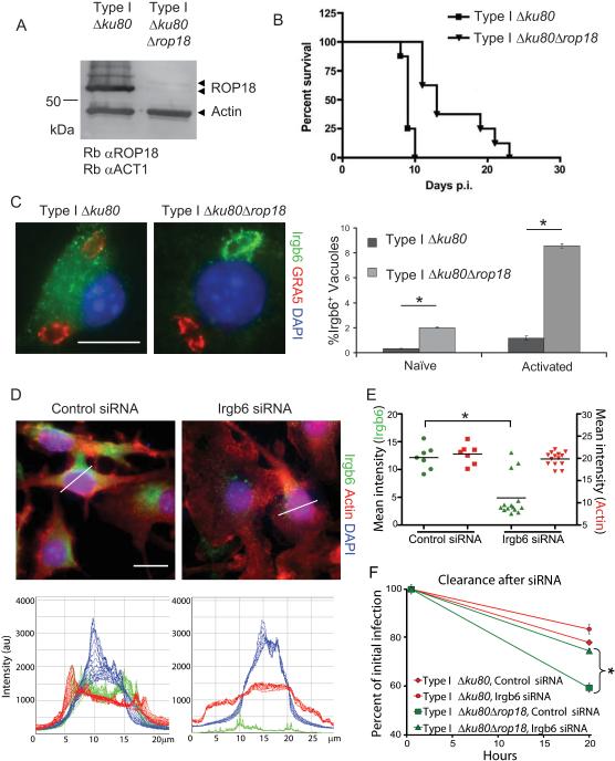
Figure 6. ROP18 is necessary to block Irgb6 recruitment.
(A) Deletion of ROP18 (Δrop18) in a Δku80 type I strain as shown by Western blotting using rabbit anti-ROP18 (Rb αROP18) and rabbit anti-actin (Rb αACT1). Faint upper band represents processing form of ROP18 (Qiu et al., 2008).
(B) Survival of mice infected by i.p. inoculation with type I Δku80 strain and Δrop18 KO. Representative of two similar experiments (P < 0.0001), n = 8 animals each.
(C) Deletion of ROP18 resulted in increased recruitment of Irgb6 in IFN-γ-activated peritoneal macrophages at 30 min post infection. Irgb6 localized with rabbit polyclonal Ab (Alexa Fluor 488, green); GRA5 localized with mAb Tg 17-113 (Alexa Fluor 594, red); and nuclei stained with DAPI (blue). Mean ± SEM, n=3 experiments (*P < 0.005, Student's t test).
(D) Immunofluorescence staining of IFN-γ-activated RAW 246.7 cells 48 h after addition of siRNAs targeting Irgb6 compared to control siRNA. Thin white lines indicate path for intensity profile analysis. Scale = 20 μm. (E) Mean intensity analysis of cells treated for RNAi. Whole cell intensities were measured using Volocity software, and mean intensities for each cell were plotted. Population mean, *P < 0.005, Mann-Whitney test. (F) Enhanced clearance of Δrop18 parasites in IFN-γ-activated RAW 264.7 macrophages was overcome by specific depletion of Irgb6 by RNAi. Mean ± SEM, n=3 experiments, *P < 0.005, Student's t test.

