
Fig. 2. SDS-PAGE analysis of purified sPBPs and their mosaics.
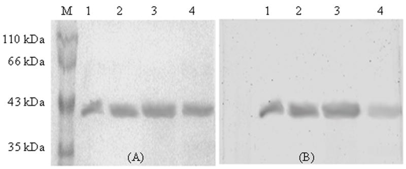
Purified sPBPs (3 μg) were stained with Coomassie blue (A), or with BOCILLIN FL (B). Lane M, Protein molecular weight markers; Lane 1, sPBP 5; Lane 2, sPBP 6; Lane 3, sPBP 656; and Lane 4, sPBP 565.