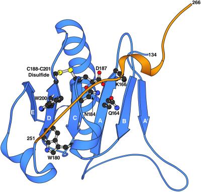
Figure 2.
Ribbon diagram representation structure of domain 2 from horse plasma gelsolin including the loop that connects S2 to S3. The other five domains are omitted for clarity. S2 is colored blue and the loop connecting S2 to S3 is orange. The strands of the β-sheet are labeled A′ to E in order from the N to C termini of the construct. The side chain of the FAF-associated mutation, residue D187, is illustrated as a ball and stick model, as are the three residues to which it is proposed to hydrogen bond in the native state (Q164, K166, N184). The two tryptophan (W180 and W200) fluorophores and the disulfide bond (C188–C201) are also shown.

