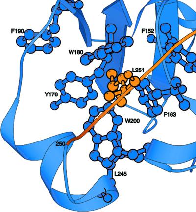
Figure 6.
Ribbon diagram representation of the hydrophobic core of horse plasma gelsolin domain 2. The other five domains and the remainder of domain 2 are omitted for clarity. The polypeptide backbone of the S2 domain (134–250) is represented in blue. The side chains of the residues that form the hydrophobic core of domain 2 are shown in blue. In orange is the polypeptide backbone of the loop connecting S2 to S3 (251–266), including the side chain of residue L251.

