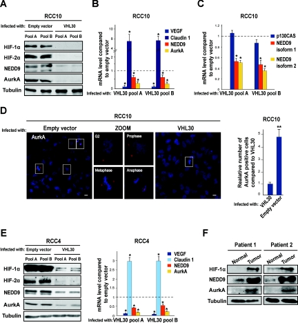
Figure 1.
Inactivation of VHL induces NEDD9 and AurkA in CCRCC cells. (A) Western blot using the indicated antibodies and lysates from two different pools of VHL-defective RCC10 cells infected with a retrovirus producing pVHL30 or control empty vector. Tubulin was used as loading control (also hereafter). (B) qPCR for the indicated targets in the same RCC10 pools; mean values of three independent experiments and the SD are shown (this applies hereafter to all data containing error bars). *P < 0.05 as analyzed by t test (also applies hereafter). (C) qPCR for NEDD9 isoforms 1 and 2 and the NEDD9-related molecule p130CAS in the same RCC10 pools. (D) Left: immunofluorescence photographs of VHL-defective RCC10 pools infected with empty vector or VHL30 and labeled for AurkA. Magnified images of the same field (only for VHL-defective cells) show AurkA staining cells at different stages of the cell cycle. Scale bars correspond to 10 μm. Right: quantification of AurkA positive cells in three immunofluorescence experiments (performed in triplicate and counting 10 fields on each coverslip) in VHL-defective RCC10 pools infected with retroviruses producing pVHL30 or empty vector. **P < 0.01 (also hereafter). (E) Western blot using the indicated antibodies (left) and qPCR for the indicated genes (right); two different pools of VHL-defective RCC4 cells were infected as above. (F) Western blot using the indicated antibodies and lysates from paired CCRCC tumor samples and surrounding normal tissue of two patients with VHL-defective sporadic CCRCC.

